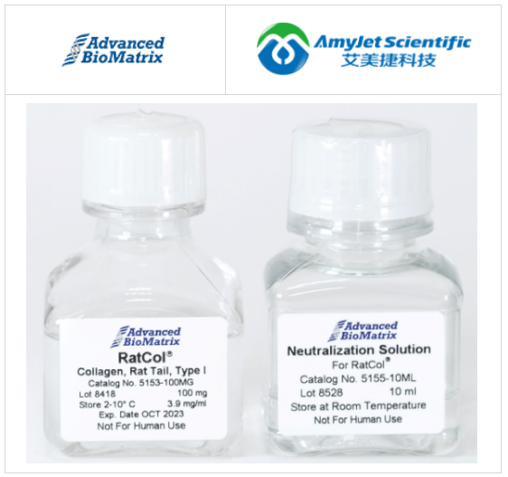
艾美捷 Advanced BioMatrixRatCol胶原蛋白溶液是一种高纯度的鼠尾胶原蛋白(100毫克),浓度约为4毫克/毫升,pH值为3-4,并经过无菌过滤。RatCol中约有97%的I型胶原蛋白,其余部分由III型胶原蛋白组成。RatCol胶原蛋白的纯度≥99%。SDS-PAGE电泳显示出典型的、和带状图案,符合胶原蛋白的特征。产品标签和每个特定批次的分析证书上打印了实际的胶原蛋白浓度。胶原蛋白附带了一瓶预配的中和溶液,用于制备胶原凝胶。RatCol胶原蛋白是通过酸提取过程从鼠尾的胶原蛋白中获得的,得到的是保持无胶原蛋白脱肽的I型胶原蛋白,溶解在0.02M乙酸缓冲液中。胶原链的N-和C末端的前胶原肽区域得以保留。I型胶原蛋白是皮肤、骨骼、肌腱和其他纤维结缔组织的主要结构成分,其与其他胶原蛋白的区别在于赖氨酸羟化和低碳水化合物含量较低。尽管已经鉴定出多种类型的胶原蛋白,但它们都由三肽链组成,呈三螺旋构象。主要结构(氨基酸序列)的轻微差异确定了不同类型之间的区别。主要结构的氨基酸序列主要是以甘氨酸为每三个位置的重复基序,脯氨酸或4-羟基脯氨酸经常位于甘氨酸残基之前。1,2 I型胶原蛋白是由两个1(I)链和一个2(I)链组成的异源三聚体,在中性pH和37C下自发形成三螺旋支架。多细胞生物中细胞生长、分化和细胞凋亡的控制依赖于细胞与细胞外基质(ECM)的黏附。

鉴于I型胶原蛋白是ECM的丰富组分,细胞在三维胶原凝胶中培养比传统的二维系统更好地模拟体内细胞环境。这已经在多种细胞类型中得到证实,包括心脏和角膜成纤维细胞、肝星状细胞(HSCs)和神经母细胞瘤细胞。3-6一些疾病可以影响ECM的力学特性,而其他疾病状态可能是由于ECM的密度或刚度变化引起的。由于I型胶原蛋白是ECM的拉伸特性的关键决定因素,三维胶原凝胶在机械转导、将机械信号转化为生化信号的细胞信号传导研究中非常有用。6-9 三维凝胶可以研究ECM的力学特性(如密度和刚度)对细胞发育、迁移和形态的影响。与二维系统不同,三维环境允许细胞伸展与所有细胞表面的整合素同时相互作用,从而激活特定的信号传导途径。凝胶的刚度或刚性在三维与二维环境中对细胞迁移产生不同的影响。
此外,3D系统中可能存在与整合素无关的机械相互作用,这是由于细胞伸展与基质纤维的纠缠而产生的,而在细胞附着于平坦表面的二维系统中则不可能发生。10-12细胞表面的一组结构和功能多样的受体识别胶原三螺旋,从而识别不同的胶原亚型。最知名的胶原受体是整合素11和21。11是平滑肌细胞上的主要整合素,而21是上皮细胞和血小板上的主要形式。这两种形式在许多细胞类型上表达,包括成纤维细胞、内皮细胞、成骨细胞、软骨细胞和淋巴细胞。13-15某些细胞类型还可能表达其他胶原受体,如糖蛋白VI(GPVI),它在血小板中介导粘附和信号传导。16其他胶原受体包括圆盘结构域受体、白细胞相关的IG样受体-1和曼诺糖受体家族成员。17,18本产品是从鼠尾腱提取的胶原蛋白制备而成,含有较高的单体含量。起始材料来自管理的斯普拉格-道利大鼠群体,并使用符合cGMP的制造过程进行纯化。该过程包含内置的、经验证的步骤,以确保可能的朊病毒和/或病毒污染物的灭活。该产品非常适用于表面涂层,提供薄层制备用于细胞培养,或作为固态凝胶使用。
文献参考:
1. Tanzer, M. L., Cross-linking of collagen. Science,180(86), 561-566 (1973).
2. Bornstein, P., and Sage, H., Structurally distinct collagen types. Ann. Rev. Biochem., 49, 957-1003 (1980).
3. Tomasek, J.J., and Hay, E.D., Analysis of the role of microfilaments in acquisition and bipolarity and elongation of fibroblasts in hydrated collagen gels. J. Cell Biol., 99, 536-549 (1984).
4. Karamichos, D., et al., Regulation of corneal fibroblast morphology and collagen reorganization by extracellular matrix mechanical properties. Invest.Ophthalmol. Vis. Sci., 48, 5030-5037(2007).
5. Sato, M., et al., 3-D Structure of extracellular matrixregulates gene expression in cultured hepaticstellate cells to induce process elongation. CompHepatol., Jan 14;3 Suppl 1:S4 (2004).
6. Li, G.N., et al., Genomic and morphologicalchanges in neuroblastoma cells in response tothree-dimensional matrices. Tissue Eng., 13, 1035-1047 (2007).
7. Roeder, B.A., et al., Tensile mechanical propertiesof three-dimensional type I collagen extracellularmatrices with varied microstructure. J. Biomech.Eng., 124, 214-222 (2002).
8. Wozniak, M.A., and Keely, P.J., Use of three-dimensionalcollagen gels to studymechanotransduction in T47D breast epithelialcells. Biol. Proced. Online, 7,144-161 (2005).
9. Grinnell, F., Fibroblast biology in three-dimensionalcollagen matrices. Trends Cell Biol., 13, 264-269(2003).
10. Beningo, K.A., et al., Responses of fibroblasts toanchorage of dorsal extracellular matrix receptors.Proc. Natl. Acad Sci. USA, 101, 18024-18029(2004).
11. Zaman, M.H., et al., Migration of tumor cells in 3Dmatrices is governed by matrix stiffness along withcell-matrix adhesion and proteolysis. Proc. Natl.Acad. Sci. USA, 103, 10889-10894 (2006).
12. Jiang, H., and Grinnell, F., Cell-matrixentanglement and mechanical anchorage offibroblasts in three-dimensional collagen matrices.Mol. Biol. Cell, 16, 5070-5076 (2005).
13. Heino, J., The collagen receptor integrins havedistinct ligand recognition and signaling functions.Matrix Biol., 19, 319-323 (2000).
14. Heino, J., The collagen family members as celladhesion proteins. BioEssays, 29, 1001-1010(2007).
15. Ivaska, J., et al., Cell adhesion to collagen-is onecollagen receptor different from another? Conn.Tiss., 30, 273-283 (1998).
16. Clemetson, K.J., and Clemetson, J.M., Plateletcollagen receptors. Thromb Haemost., 86, 189-197(2001).
17. Leitinger, B., and Hohenester,E., MammalianCollagen Receptors, Matrix Biol., 26, 146-155(2007).
18. Popova, S.N., et al., Physiology and pathology ofcollagen receptors. Acta Physiol. (Oxf), 190, 179-187 (2007).
本文作者可以追加内容哦 !

